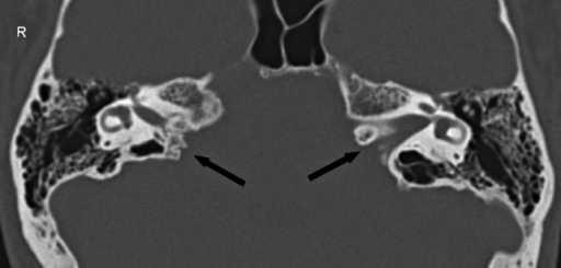

The internal auditory canal (IAC) is situated at the posterior aspect of the petrous part of the temporal bone.

Arnau Benet, in Handbook of Clinical Neurology, 2020 Dural reflections at the region of the internal auditory canal The literature also describes rare cases of bilateral, duplicated IACs that were associated with labyrinthine anomalies. 55 After cochlear implantation, some patients with narrow IACs have experienced facial pain and twitching without useful auditory sensation. 143 A narrow IAC has been considered a relative contraindication to cochlear implantation, because it suggests that the eighth cranial nerve may be insufficiently developed to conduct an auditory signal. Interestingly, in patients with atresia of the IAC, the facial nerve may take an aberrant course to establish facial motor function. A narrow IAC may accompany inner ear malformations or may be the sole radiographically detectable anomaly in a deaf child. When a patient has normal facial function and an IAC less than 3 mm in diameter, it is likely that the bony canal transmits only the facial nerve ( Fig. Jackler, in Cummings Pediatric Otolaryngology (Second Edition), 2021 Narrow Internal Auditory CanalĪ narrow IAC may indicate a failure of eighth cranial nerve development. Intraosseous schwannoma of the petrous apex. Goiney C, Bhatia R, Auerbach K, Norenberg M, Morcos J. The Natural History and Management of Petrous Apex Cholesterol Granulomas. Sweeney AD, Osetinsky LM, Carlson ML, et al. Cholesterol Granulomas: A Comparative Meta-Analysis of Endonasal Endoscopic versus Open Approaches to the Petrous Apex. The preauricular subtemporal approach for transcranial petrous apex tumors. Leonetti JP, Anderson DE, Marzo SJ, Origitano TC, Schuman R. Surgical outcomes after endoscopic management of cholesterol granulomas of the petrous apex: a systematic review. 80 (5):500-4.Įytan DF, Kshettry VR, Sindwani R, et al. Novel Application of Steroid Eluting Stent in Petrous Apex Cholesterol Granuloma. 104(1):29-36.Ĭhoi KJ, Jang DW, Zomorodi AR, Codd PJ, Friedman A, Abi Hachem R. Transcanal infracochlear approach to the petrous apex. Endoscopic endonasal and transorbital approaches to petrous apex lesions. Landmarks to Identify Petrous Apex Through Endonasal Approach Without Transgression of Sinus. Negm HM, Singh H, Dhandapani S, Cohen S, Anand VK, Schwartz TH. Superior sagittal sinus thrombosis after radical neck dissection. Cystic lesions of the petrous apex: transphenoidal approach. Subtemporal transpetrosal apex approach: study on its use in large and giant petroclival meningiomas. Yang J, Ma SC, Fang T, Qi JF, Hu YS, Yu CJ. Neurosurgical strategies and operative results in the treatment of tumors of or extending to the petrous apex. Radiographic differential diagnosis of petrous apex lesions. Infralabyrinthine approach to the petrous apex. Pneumatization Patterns of the Petrous Apex and Lateral Sphenoid Recess. Malone A, Bruni M, Wong R, Tabor M, Boyev KP. Petrous pyramid of temporal bone: Pneumatization and roentgenologic appearance. Sulla leptominingite circiscritta e sulla paralisi dell' abducente di origine otitica. Li KL, Agarwal V, Moskowitz HS, Abuzeid WM.


 0 kommentar(er)
